Fundamentals of SEM In principle the Scanning Electron Microscope allows us to visualize material by the scanning of a fine beam of electrons over the surface of a specimen in synchronism with the spot of the display cathode ray tube (CRT). Electrons can be emitted from a tungsten cathode or via field emission A detector monitors the intensity of a secondary signal from the specimen (e,g, secondary electrons)and the brightness of the CRT spot is controlled by an amplified version of the detected signal.
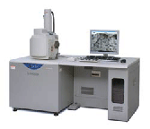
Hitachi S-3400N SEM is a high performance, user-friendly scanning electron microscope with new improvements that allow the best results for a wide range of applications. It is equipped with both Secondary (SE) and Backscatter (BSE) Electron Detectors and has a fully eucentric 5 axis motorized stage that allows samples up to 25cm in diameter. The Variable Pressure mode allows BSE observation from 6- 270Pa, and the instrument is equipped with a Deben Peltier Coolstage to control moisture loss in low vacuum mode.
Specifications
Range : up to 300000x


